Anti-PARP antibody
| 英文名称 | PARP |
| 中文名称 | 多腺苷二磷酸多聚酶抗体(N端) |
| 别 名 | ADP ribosyltransferase (NAD+; poly (ADP ribose) polymerase); ADP ribosyltransferase NAD+; ADPRT 1; ADPRT; ADPRT1; msPARP; NAD(+) ADP ribosyltransferase 1; pADPRT 1; pADPRT1; PARP 1; PARP1; Poly (ADP ribose) polymerase 1; poly (ADP ribose) polymerase family, member 1; Poly adenosine diphosphate ADP ribose polymerase; Poly ADP ribose polymerase 1; Poly ADP ribose polymerase family member 1; Poly ADP ribose synthetase 1; poly(ADP ribose) synthetase; poly(ADP ribosyl)transferase; Poly[ADP ribose] synthetase 1; PPOL; sPARP 1; sPARP1; PARP1_HUMAN. |
DATASHEET
Host:Rabbit
Target Protein:PARP
IR:Immunogen Range:201-300/1014
Clonality:Polyclonal
Isotype:IgG
Entrez Gene:142
Swiss Prot:P09874
Source:KLH conjugated synthetic peptide derived from human PARP:201-300/1014
Purification:affinity purified by Protein A
Storage:0.01M TBS(pH7.4) with 1% BSA, 0.03% Proclin300 and 50% Glycerol. Shipped at 4℃. Store at -20 °C for one year. Avoid repeated freeze/thaw cycles.
Background:This gene encodes a chromatin-associated enzyme, poly(ADP-ribosyl)transferase, which modifies various nuclear proteins by poly(ADP-ribosyl)ation. The modification is dependent on DNA and is involved in the regulation of various important cellular processes such as differentiation, proliferation, and tumor transformation and also in the regulation of the molecular events involved in the recovery of cell from DNA damage. In addition, this enzyme may be the site of mutation in Fanconi anemia, and may participate in the pathophysiology of type I diabetes. [provided by RefSeq, Jul 2008].
Size:100ul
Concentration:1mg/ml
Applications:WB(1:500-2000)
ELISA(1:5000-10000)
IHC-P(1:100-500)
IHC-F(1:100-500)
Flow-Cyt(0.2μg/Test)
IF(1:100-500)
Cross Reactive Species:Human
Mouse
Rat
Dog
Cow
.
For research use only. Not intended for diagnostic or therapeutic use.
VALIDATION IMAGES
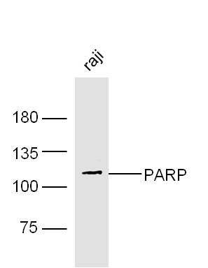
Sample:
Raji Cell (Human) Lysate at 30 ug
Primary: Anti- PARP (bs-2138R) at 1/300 dilution
Secondary: IRDye800CW Goat Anti-Rabbit IgG at 1/20000 dilution
Predicted band size: 111 kD
Observed band size: 111 kD
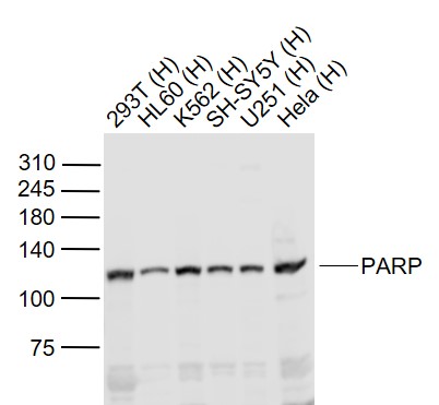
Sample:
Lane 1: 293T (Human) Cell Lysate at 30 ug
Lane 2: HL60 (Human) Cell Lysate at 30 ug
Lane 3: K562 (Human) Cell Lysate at 30 ug
Lane 4: SH-SY5Y (Human) Cell Lysate at 30 ug
Lane 5: U251 (Human) Cell Lysate at 30 ug
Lane 6: Hela (Human) Cell Lysate at 30 ug
Primary: Anti-PARP (bs-2138R) at 1/1000 dilution
Secondary: IRDye800CW Goat Anti-Rabbit IgG at 1/20000 dilution
Predicted band size: 115 kD
Observed band size: 115 kD
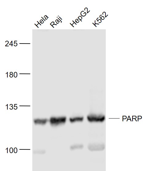
Sample:
Hela(Human) Cell Lysate at 30 ug
Raji(Human) Cell Lysate at 30 ug
HepG2(Human) Cell Lysate at 30 ug
K562(Human) Cell Lysate at 30 ug
Primary: Anti- PARP (bs-2138R) at 1/1000 dilution
Secondary: IRDye800CW Goat Anti-Rabbit IgG at 1/20000 dilution
Predicted band size: 111 kD
Observed band size: 111 kD
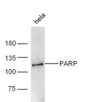
Sample:
Hela Cell (Human) Lysate at 30 ug
Primary: Anti- PARP (bs-2138R) at 1/300 dilution
Secondary: IRDye800CW Goat Anti-Rabbit IgG at 1/20000 dilution
Predicted band size: 111 kD
Observed band size: 111 kD
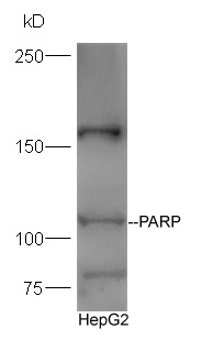
Protein:HepG2 lysate at 30ug;
Primary: Anti-PARP (bs-2138R) at 1:300 dilution;
Secondary: HRP conjugated Goat-Anti-rabbit IgG(bs-0295G-HRP) at 1: 5000 dilution;
Predicted band size:111 kD
Observed band size:111 kD
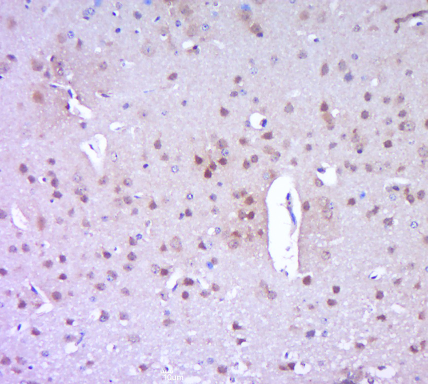
Paraformaldehyde-fixed, paraffin embedded (mouse brain tissue); Antigen retrieval by boiling in sodium citrate buffer (pH6.0) for 15min; Block endogenous peroxidase by 3% hydrogen peroxide for 20 minutes; Blocking buffer (normal goat serum) at 37°C for 30min; Antibody incubation with (PARP) Polyclonal Antibody, Unconjugated (bs-2138R) at 1:400 overnight at 4°C, followed by a conjugated secondary (sp-0023) for 20 minutes and DAB staining.
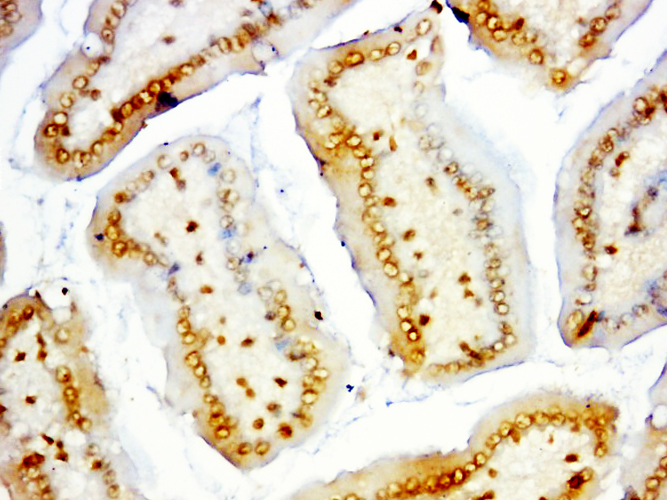
Paraformaldehyde-fixed, paraffin embedded (mouse intestines); Antigen retrieval by boiling in sodium citrate buffer (pH6.0) for 15min; Block endogenous peroxidase by 3% hydrogen peroxide for 20 minutes; Blocking buffer (normal goat serum) at 37°C for 30min; Antibody incubation with (PARP) Polyclonal Antibody, Unconjugated (bs-2138R) at 1:500 overnight at 4°C, followed by a conjugated secondary (sp-0023) for 20 minutes and DAB staining.
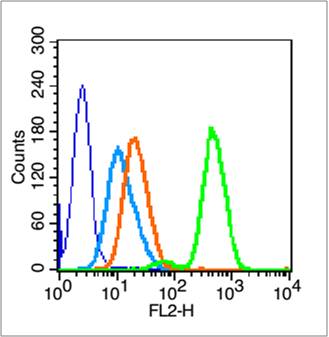
Blank control (blue line): HL60 cells (blue).
Primary Antibody (green line): Rabbit Anti- PARP antibody (bs-2138R)
Dilution: 0.2μg /10^6 cells;
Isotype Control Antibody (orange line): Rabbit IgG .
Secondary Antibody (white blue line): Goat anti-rabbit IgG-PE
Dilution: 1μg /test.
Protocol
The cells were fixed with 70% methanol (Overnight at 4℃) and then permeabilized with 90% ice-cold methanol for 20 min at -20℃. Cells stained with Primary Antibody for 30 min at room temperature. The cells were then incubated in 1 X PBS/2%BSA/10% goat serum to block non-specific protein-protein interactions followed by the antibody for 15 min at room temperature. The secondary antibody used for 40 min at room temperature. Acquisition of 20,000 events was performed.
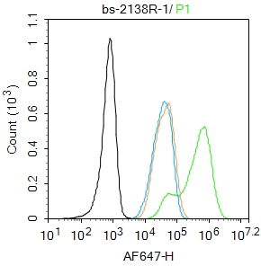
Blank control:293T.
Primary Antibody (green line): Rabbit Anti-PARP1 antibody (bs-2138R)
Dilution: 1μg /10^6 cells;
Isotype Control Antibody (orange line): Rabbit IgG .
Secondary Antibody : Goat anti-rabbit IgG-AF647
Dilution: 1μg /test.
Protocol
The cells were fixed with 4% PFA (10min at room temperature)and then permeabilized with 90% ice-cold methanol for 20 min at -20℃.The cells were then incubated in 5%BSA to block non-specific protein-protein interactions for 30 min at room temperature .Cells stained with Primary Antibody for 30 min at room temperature. The secondary antibody used for 40 min at room temperature. Acquisition of 20,000 events was performed.


好评度