Anti-Estrogen receptor alpha antibody
| 英文名称 | Estrogen receptor alpha |
| 中文名称 | 雌激素受体α抗体 |
| 别 名 | Estradiol receptor; Estradiol Receptor-alpha; Estrogen Receptor 1; Atherosclerosis, susceptibility to, included; DKFZp686N23123; ER Alpha; ER; ER-alpha; ERalpha; ER[a]; Era; ESR; ESR1; ESR1_HUMAN; ESR2; ESRA; Estr; Estrogen receptor 1 (alpha); Estrogen resistance, included; HDL cholesterol, augmented response of, to hormone replacement, included; Myocardial infarction, susceptibility to, included; NR3A1; Nuclear receptor subfamily 3 group A member 1; OTTHUMP00000017718; OTTHUMP00000017719; RNESTROR. |
DATASHEET
Host:Rabbit
Target Protein:Estrogen receptor alpha
IR:Immunogen Range:501-595/595
Clonality:Polyclonal
Isotype:IgG
Entrez Gene:2099
Swiss Prot:P03372
Source:KLH conjugated synthetic peptide derived from human ER-Alpha:501-595/595
Purification:affinity purified by Protein A
Storage:0.01M TBS(pH7.4) with 1% BSA, 0.03% Proclin300 and 50% Glycerol. Shipped at 4℃. Store at -20 °C for one year. Avoid repeated freeze/thaw cycles.
Background:Estrogen and progesterone receptor are members of a family of transcription factors that are regulated by the binding of their cognate ligands. The interaction of hormone-bound estrogen receptors with estrogen responsive elements(EREs) alters transcription of ERE-containing genes. The carboJNC terminal region of the estrgen receptor contains the ligand binding domain, the amino terminus serves as the transactivation domain, and the DNA binding domain is centrally located. Two forms of estrogen receptor have been identified, ER Alpha and ER Beta. ER Alpha and ER Beta have been shown to be differentially activated by various ligands. The biological response to progesterone is mediated by two distinct forms of the human progesterone receptor (hPR-A and hPR-B), which arise from alternative splicing. In most cells, hPR-B functions as a transcriptional activator of progesterone-responsive gene, whereas hPR-A function as a transcriptional inhibitor of all steroid hormone receptors.
Size:100ul
Concentration:1mg/ml
Applications:WB(1:500-2000)
ELISA(1:5000-10000)
Flow-Cyt(1μg/Test)
ICC(1:100)
Cross Reactive Species:Human
Mouse
.
For research use only. Not intended for diagnostic or therapeutic use.
VALIDATION IMAGES
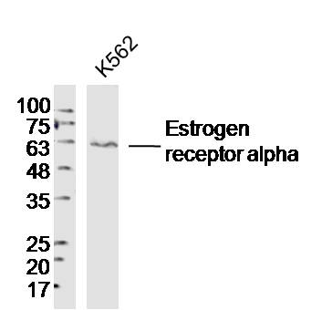
Sample:K562 (Human)Cell Lysate at 40 ug
Primary: Anti-Estrogen receptor alpha(bs-0254R)at 1/300 dilution
Secondary: IRDye800CW Goat Anti-RabbitIgG at 1/20000 dilution
Predicted band size: 66kD
Observed band size: 63kD
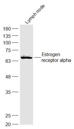
Sample:
Lymph node (Mouse) Lysate at 40 ug
Primary: Anti-Estrogen receptor alpha (bs-0254R) at 1/300 dilution
Secondary: IRDye800CW Goat Anti-Rabbit IgG at 1/20000 dilution
Predicted band size: 66 kD
Observed band size: 66 kD
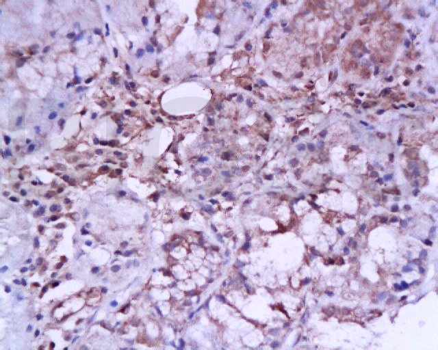
Tissue/cell: Human laryngeal carcinoma; 4% Paraformaldehyde-fixed and paraffin-embedded;
Antigen retrieval: citrate buffer ( 0.01M, pH 6.0 ), Boiling bathing for 15min; Block endogenous peroxidase by 3% Hydrogen peroxide for 30min; Blocking buffer (normal goat serum,C-0005) at 37℃ for 20 min;
Incubation: Anti-ER-alpha Polyclonal Antibody, Unconjugated(bs-0254R) 1:200, overnight at 4°C, followed by conjugation to the secondary antibody(SP-0023) and DAB(C-0010) staining
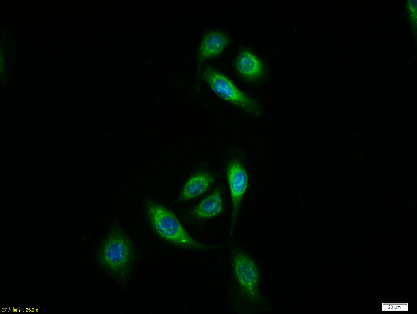
Tissue/cell:MCF7 cell; 4% Paraformaldehyde-fixed; Triton X-100 at room temperature for 20 min; Blocking buffer (normal goat serum, C-0005) at 37°C for 20 min; Antibody incubation with (Estrogen receptor alpha) polyclonal Antibody, Unconjugated (bs-0254R) 1:100, 90 minutes at 37°C; followed by a FITC conjugated Goat Anti-Rabbit IgG antibody at 37°C for 90 minutes, DAPI (blue, C02-04002) was used to stain the cell nuclei.


好评度